
Transitional Epithelium Definition
Transitional epithelium is a stratified tissue made of multiple cell layers, where the cells constituting the tissue can change shape depending on the distention in the organ. When the organ is filled with fluid, cells on the topmost layer of this epithelium can stretch and appear flattened. Alternately, they can also appear cuboidal with a rounded shape when the fluid pressure is low.
Function of Transitional Epithelium
Due to its location in the excretory system, especially in the ureters and urinary bladder, one of the primary functions of this tissue is to be an extremely effective permeability barrier, impenetrable to water and most small molecules. The cells of this tissue are probably among the most resistant to osmotic pressure.
Urine is hypertonic with a much higher concentration of many solutes compared to the cytoplasm of the epithelial cells. Yet, these cells are protected from desiccation even when the epithelium is fully stretched. Toxic wastes are also prevented from re-entering the blood stream.
The second important function of these cells is to allow the organ to stretch and increase its volume depending on fluid pressure. For instance, when there is a lot of water that needs to be expelled from the body, a large quantity of liquid passes through the ureters, urinary bladder and urethra.
The ability of the cells in the superficial layer of this epithelium to change shape (transition from a rounded cuboidal shape to a flattened squamous structure) allows these organs to stretch without exposing underlying tissue to the toxic substances in the urine.
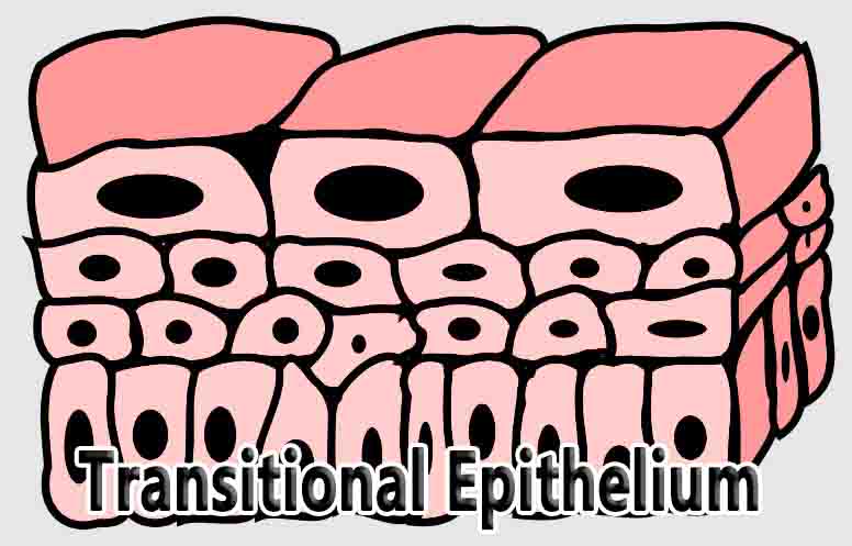
Structure of Transitional Epithelium
Transitional epithelia are made of 3-4 layers of cells with the lowermost or basal layer staying in contact with the basement membrane. The cells of this basal layer are attached to the lamina propria through tonofilaments and hemi-desmosomes. These are among the least differentiated cells in this tissue and support the remaining cells. Cells in the intermediate layers are proliferative and can replenish cells lost due to abrasion or infection.
They also have a vast Golgi network that contains a number of membrane-bound vesicles. The superficial layer of cells can change from being cuboidal to appearing flattened when the organ is distended and contain a number of actin-based cytoplasmic projections known as microvilli. A two-dimensional network of hexameric plaques covers the apical plasma membrane of these cells.
These plaques are made from a protein called uroplakin and are an important feature of this epithelium, contributing to the permeability barrier for water, ammonia, urea and many other solutes of the urine. They are also likely to be involved in the ability of these cells to transition in shape.
All the cells of this epithelium are deeply connected to each other through junctional complexes. Junctional complexes are symmetrical attachments between two cells usually made of three components: a band of tight junctions on the apical surface, followed by an intermediate series of adherens junctions and basally located desmosomes.
Together, these multiprotein complexes hold the cells of the epithelium together and present an uninterrupted surface to the lumen of the organ.
Examples of Transitional Epithelium
Transitional epithelia are most commonly found in the urinary and male reproductive tract in humans. These are regions where the volume and osmolarity of the organ can change rapidly. In the urinary system, the volume and concentration of solutes in the urine depends on a number of factors.
Similarly, the prostatic urethra in the male reproductive system is lined by transitional epithelium continuous with the epithelium of the bladder. It is the most dilatable part of the urethra, expanding or contracting depending on the flow of urine or semen.
Bladder
The bladder is an organ that is designed to hold a large proportion of the body’s toxic, liquid waste, before it is expelled from the body. When fully distended, the urinary bladder can hold nearly 500 ml of urine, making it an organ that has drastic changes in volume over short spans of time.
While three layers of muscle fibers contribute towards the distention and contraction of the organ, the transitional epithelium is also crucial. The junctional complexes and uroplakin plaques of the superficial cells protect the body from the effects of storing urea, ammonia and other metabolites in the bladder.
In addition, the plaques are said to help apical cells adjust the surface area of their plasma membrane. For instance, when the bladder is distended, there is an increase in the membrane area, possibly through the fusion of vesicles from the Golgi network.
Related Biology Terms
- Adherens Junctions – Protein complexes that constitute a part of cell-cell adhesions, which involve the actin cytoskeleton. They lie basal to tight junctions and can encircle the cell. Also known as zonula adherens.
- Desmosomes – Spot-like adhesions between two cells made of cadherin proteins that allow cells in a tissue to resist shearing forces. Also known as macula adherens.
- Lamina Propria – Thin layer of connective tissue that lies underneath epithelia, constituting one part of the mucosa. Mucosa lines various cavities in the body and surrounds organs.
- Tonofibrils – Intermediate filaments made of keratin, which converge at desmosomes and hemidesmosomes, attaching the cell to other cells or the extracellular matrix.