
Definition
At the base of the mastoid area of the temporal bone, the mastoid process is a smooth conical projection of bone. Among the muscles attached to it are the occipitofrontalis and sternocleidomastoid muscles, as well as the splenius capitis muscle.
Mastoid Process Location
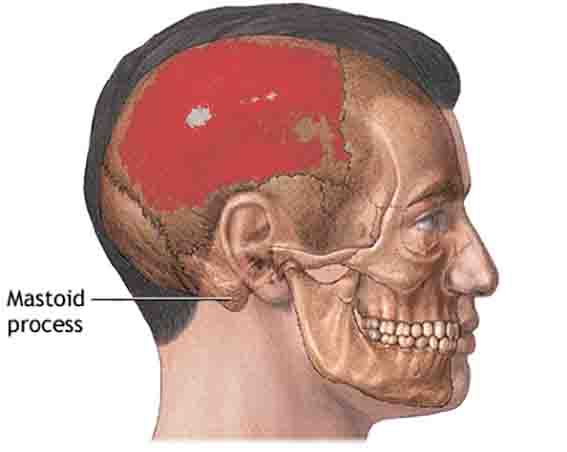
On the underside of the mastoid portion of the temporal bone, behind the external auditory meatus, is the mastoid process. In front of and behind the ear canal and lateral to the styloid process, it can be found.
The petrosquamous suture travels vertically from the superior border of the mastoid area to the parietal bone. As the anterior border articulates with the ascending part of the temporal bone’s squamous area, the posterior border merges with the occipital bone.
Mastoid Process Anatomy
The mastoid process is a smooth, conical projection of bone with several structures that allow it to perform its specific functions. Among them are:
- On the medial side of the mastoid process is the digastric fossa/mastoid notch
- Located medial to the digastric fossa, the occiput groove
- A mastoid air cell/Lenoir is a hollow area within the mastoid process located in its superior, inferior, and anterior areas. Superior and anterior parts are larger and more irregular, while inferior parts are smaller
Mastoid Process Function
The mastoid process provides an area of attachment to several important head muscles. It is the attachment site for certain neck muscles, such as:
- Rotation of the head to the contralateral side is enabled by the Sternocleidomastoid muscle
- Head extension, rotation, and lateral flexion are controlled by the Splenius capitis muscle
- When the masseter and temporalis muscles relax, the posterior belly of the digastric muscle opens the jaw.
- The longissimus capitis muscle is responsible for head lateral rotation
From the superior nuchal line to the mastoid process, the occipitofrontalis muscle anchors on the rough outer surface of the mastoid process.
Additionally, the digastric fossa is where the digastric muscle attaches, while the occipital artery attaches to the medial occipital groove.
The mastoid air cells present in the mastoid process also serve their own functions, including protecting the temporal bone and the middle and inner ear from trauma, and regulating air pressure. Aditus ad antrum and mastoid antrum connect them with the middle ear.
Mastoid Process Disease
A mastoid process can be affected by one or more diseases, just as any other part of the body. The mastoid process can be affected by a variety of conditions, such as:
Mastoiditis
Otitis media (middle ear infection) is a fairly common condition, particularly in children, and is often easily treatable. Untreated, aditus ad antrum and mastoid antrum can spread to the mastoid area, leading to mastoiditis.
Mastoiditis is an infection of the mastoid antrum mucus lining and the mastoid air cells in the mastoid process. Mastoiditis is characterized by the following symptoms:
- Pain or swelling behind the ear in the mastoid process area
- Discharge from the ears
- The fever
- Having a headache
- Loss of hearing
Bacteria can cause mastoiditis in several different ways, the most common being:
- The pneumonia-causing bacteria Streptococcus pneumoniae
- It is caused by a bacteria called Streptococcus pyogenes (also known as scarlet fever bacteria).
- A streptococcal infection
- Influenza virus (Haemophilus influenzae)
- The moraxella catarrhalis bacteria
Mastoiditis can lead to a variety of complications if left untreated:
- Mastoid process destruction
- Infection with meningitis
- Loss of hearing in part or in full
- Infection of the brain
Despite the fact that mastoiditis is often located deep within the bone, it can be treated with antibiotics, either injected or taken orally (or sometimes both). In the event that this does not work, mastoidectomy can be performed to remove part of the mastoid process and drain it.
Cholesteatoma
Cholesteatomas are abnormal growths of skin cells in the middle ear or mastoid process. A history of repeated middle ear infections is more likely to cause cholesteatoma than a birth defect. Despite not being cancerous, this growth can still cause a number of issues, including:
- Ear discharge that is watery
- In the affected ear, there is a gradual loss of hearing
- Pressure in the ear that is uncomfortable
As the cholesteatoma cyst grows over time, these symptoms will gradually worsen, and if left untreated they will worsen. A number of unpleasant complications can result from this, including:
- An ear infection
- Loss of hearing
- Hearing loss
- Nerve damage to the face
- An abscessed brain
- The meningitis virus
It is therefore crucial to catch and treat cholesteatoma as soon as possible. During surgery, the cholesteatoma is permanently removed while the patient is under general anesthesia. In addition to antibiotics, antibiotics may also be prescribed to combat infections in the cyst or ear, making it easier to observe the cyst’s growth traits to plan its surgical removal.